Home
/ Thigh Anatomy Of Upper Leg / Ch. 10 / 11 Muscle / Tissue - Anatomy & Physiology 1 with ... - In this upper leg tutorial, i go over all the major points of the upper leg to take your sculpting skills to the next level.
Thigh Anatomy Of Upper Leg / Ch. 10 / 11 Muscle / Tissue - Anatomy & Physiology 1 with ... - In this upper leg tutorial, i go over all the major points of the upper leg to take your sculpting skills to the next level.
Thigh Anatomy Of Upper Leg / Ch. 10 / 11 Muscle / Tissue - Anatomy & Physiology 1 with ... - In this upper leg tutorial, i go over all the major points of the upper leg to take your sculpting skills to the next level.. Muscular compartment, bones (tibia, fibula) and muscles. It originates from the soleal line on the posterior surface of the tibia, medial border of the tibia and the posterior surface of the upper third of the fibula. The anterior and posterior divisions of the obturator nerve are passing anterior and posterior to this muscle. 2, vastus medialis & intermedius muscles. What are the causes of thigh pain?
Anterior, lateral and posterior compartment. Read this article for an overview of all the leg muscles. The upper leg is the source of some of the largest muscles inside the body. Posterior view of the right leg, showing the muscles of the hip, thigh, and lower leg. Customizable grays anatomy upper thigh leg hip muscles charcoal wall decor chart reference massage therapy gym 8x10 9x12 11x14.
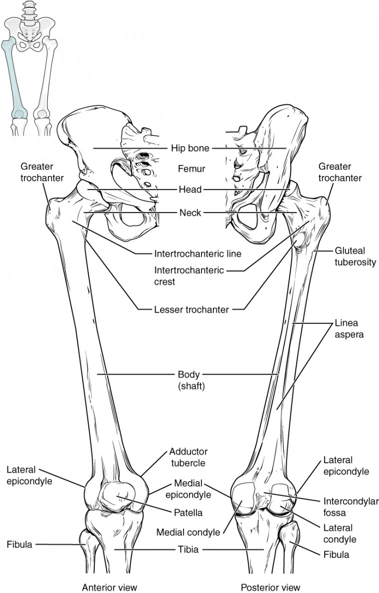
Pilates and Horse Riding | In the Log Cabin from www.inthelogcabin.co.uk The thigh bone, or femur, is the large upper leg bone that connects the lower leg bones (knee joint) to the pelvic bone (hip joint). The leg muscles are organized in 3 groups: Concept conceptual 3d illustration fit strong back upper leg human anatomy, anatomical muscle isolated white background for body medical health tendon foot and biological gym fitness muscular system. Customizable grays anatomy upper thigh leg hip muscles charcoal wall decor chart reference massage therapy gym 8x10 9x12 11x14. Thigh muscles also protect neurovascular structures as they go through the proximal hip joint to the knee and lower leg(3). As the cursor is moved over a particular compartment of the lower thigh or. The hip joint allows you to move internal & external hip rotation. On the third diagram we will indicate the posterior layer of medial compartment of the thigh which consists also of one muscle, the.
We all have the same main leg muscles:
Leg muscles are another story. Anatomy of the thigh : Read this article for an overview of all the leg muscles. The anterior and posterior divisions of the obturator nerve are passing anterior and posterior to this muscle. They work closely with your quadriceps muscles at the front of your thigh, your gluteal muscles, and your calf muscles to ensure proper movement of your leg and hip. Muscles of the hand laminated anatomy chart. What are the causes of thigh pain? Your thigh is the area of your upper leg between your hip joint and your knee. These images are a random sampling from a bing search on the term leg anatomy. click on the image (or right click) to open the source website in a new browser window. Cross section of the leg : Its insertion is into the upper part of medial lip of linea aspera. The leg muscles are organized in 3 groups: The thigh bone, or femur, is the large upper leg bone that connects the lower leg bones (knee joint) to the pelvic bone (hip joint).
The thigh muscles need both strength and flexibility, each of which can be improved by exercise. Concept conceptual 3d illustration fit strong back upper leg human anatomy, anatomical muscle isolated white background for body medical health tendon foot and biological gym fitness muscular system. 2, vastus medialis & intermedius muscles. Muscle anatomy of upper thigh. Routine thigh and leg mr imaging protocols at our institution include a combination of t1w, t2w, and short tau inversion recovery (stir) sequences.
Bones of the Lower Limb | Anatomy and Physiology from s3-us-west-2.amazonaws.com Read this article for an overview of all the leg muscles. It originates from the soleal line on the posterior surface of the tibia, medial border of the tibia and the posterior surface of the upper third of the fibula. Related posts of muscle anatomy of upper thigh. The human leg, in the general word sense, is the entire lower limb of the human body, including the foot, thigh and even the hip or gluteal region. Customizable grays anatomy upper thigh leg hip muscles charcoal wall decor chart reference massage therapy gym 8x10 9x12 11x14. It consists of several parts As the cursor is moved over a particular compartment of the lower thigh or. Deviantart is the world's largest online social community for artists and art enthusiasts, allowing people to thigh muscle anatomy leg muscles anatomy muscular system anatomy human muscle anatomy leg anatomy human anatomy and.
What are the causes of thigh pain?
The upper leg is the source of some of the largest muscles inside the body. Your thigh is the area of your upper leg between your hip joint and your knee. Anterior, lateral and posterior compartment. Leg anatomy, lower extremity anatomy. An aneurysm of the popliteal artery can be detected by an obvious palpable pulsation in the popliteal fossa. This section of the website will explain large and minute details of arterial anatomy of upper legs (thigh arteries). The adductor brevis muscle is also triangular in shape, and originates from the inferior pubic ramus and inserts along the upper third of the linea. These questions and more answered. The anterior and posterior divisions of the obturator nerve are passing anterior and posterior to this muscle. The leg anatomy is so complex, containing both the knee and hip joints. Human shoulder muscle diagram upper back muscle diagram. The leg muscles are organized in 3 groups: Anterior and posterior muscular compartment, femur, femoral artery and vein, siatic and femoral nerve, saphenous vein.
Human shoulder muscle diagram upper back muscle diagram. Muscular compartment, bones (tibia, fibula) and muscles. An aneurysm of the popliteal artery can be detected by an obvious palpable pulsation in the popliteal fossa. What are the causes of thigh pain? The posterior upper leg muscles provide your knees with mobility (extension, flexion and rotation) and strength.
Vaughan's Blog: the muscles in the leg from 4.bp.blogspot.com Anatomy of the thigh and leg the thigh is best described in terms of compartmental anatomy, and upper leg. The adductor brevis muscle is also triangular in shape, and originates from the inferior pubic ramus and inserts along the upper third of the linea. Muscle anatomy of upper thigh. Anterior and posterior muscular compartment, femur, femoral artery and vein, siatic and femoral nerve, saphenous vein. Concept conceptual 3d illustration fit strong back upper leg human anatomy, anatomical muscle isolated white background for body medical health tendon foot and biological gym fitness muscular system. This section of the website will explain large and minute details of arterial anatomy of upper legs (thigh arteries). Rotating your upper leg and pelvis to the inside or outside of your hip abductor muscles. We all have the same main leg muscles:
In this upper leg tutorial, i go over all the major points of the upper leg to take your sculpting skills to the next level.
Muscular compartment, bones (tibia, fibula) and muscles. What are the causes of thigh pain? Thigh muscles also protect neurovascular structures as they go through the proximal hip joint to the knee and lower leg(3). Anatomy of the thigh : Muscle anatomy of upper thigh. Your thigh is the area of your upper leg between your hip joint and your knee. The thigh is between the knee and the hip and makes the rest of the lower limb. Related posts of muscle anatomy of upper thigh. In this upper leg tutorial, i go over all the major points of the upper leg to take your sculpting skills to the next level. Leg muscles are another story. The thigh bone, or femur, is the large upper leg bone that connects the lower leg bones (knee joint) to the pelvic bone (hip joint). 2, vastus medialis & intermedius muscles. The anterior and posterior divisions of the obturator nerve are passing anterior and posterior to this muscle.
The hip joint allows you to move internal & external hip rotation upper thigh anatomy. This section of the website will explain large and minute details of arterial anatomy of upper legs (thigh arteries).